PR APP, Breast Cancer PR APP, Breast Cancer

Process APP

Revert

#90003

CE-IVD

PR APP, Breast Cancer

Certified under IVDR

According to immunohistochemistry (IHC), progesteron receptor (PR) is used to determine prognosis and as a predictive marker in breast cancer. The American Society of Clinical Oncology/College of American Pathologists (ASCO/CAP) recommends that the PR status of patients is determined on all invasive breast cancers and breast cancer recurrences[1]. It is, additionally, recommended that the PR status of the tumor is considered positive if there are at least 1 % positive tumor nuclei in a sample.

This protocol can be used to determine PR positivity and negativity in a tumor, and provides the number of positive nuclei as well as the total number of nuclei. In addition, the ratio of positive nuclei and the area ratio of positive nuclei are given. Tumor regions must be identified and outlined manually within a region of interest (ROI). Calibration of the protocol allows it to be used on images with different staining intensities.

In EU/UK: CE IVD – for use in diagnostic procedures

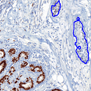
Figure 1

Figure 1

Manual outline of a tumor region.

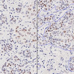
Figure 2

Figure 2

The same field of view as in FIGURE 1 after detection of nuclei as either PR-positive (red label) or PR-negative (blue label). From this classification, the output variables can be calculated.

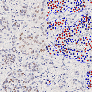
Figure 3

Figure 3

A field of view including an un-biased counting frame, ensuring that nuclei are counted only once.

Details

Quantitative Output variables

The primary outputs of the device are:

Positive Percentage,
which is a value between 0 and 100 and is calculated as

Positive Percentage = (Number of ER positive tumor nuclei)/(Total number of tumor nuclei) × 100

Allred Score,
which is a value between 0 and 8 and is calculated as

Allred Score = Proportion Score + Intensity Score

where Proportion Score is a value between 0 and 5 which reflects the positive percentage, and Intensity Score is a value between 0 and 3 which reflects the intensity of the ER positive nuclei.

The secondary outputs of the device are:

  • Neg Nuclei (#), which is the number of tumor nuclei negative for ER
  • Pos Nuclei (#), which is the number of tumor nuclei positive for ER
  • Total Nuclei (#), which is the total number of tumor nuclei
  • Allred Proportion Score
  • Allred Intensity Score

Workflow

Step 1: Manually outline tumor areas as regions of interest (ROIs)

Step 2: Load and run the 90003 PR APP, Breast Cancer to analyze the nuclei in tumor regions

References

1. Allison KH, Hammond MEH, Dowsett M, McKernin SE, Carey LA, Fitzgibbons PL, Hayes DF, Lakhani SR, Chavez-MacGregor M, Perlmutter J, Perou CM, Regan MM, Rimm DL, Symmans WF, Torlakovic EE, Varella L, Viale G, Weisberg TF, McShane LM, Wolff AC. Estrogen and Progesterone Receptor Testing in Breast Cancer: ASCO/CAP Guideline Update. J Clin Oncol. 2020 Apr 20;38(12):1346-1366.


Success
Your message has been successfully sent!