#10150
Picrosirius Red, SADS
Developed for St. George’s University of London
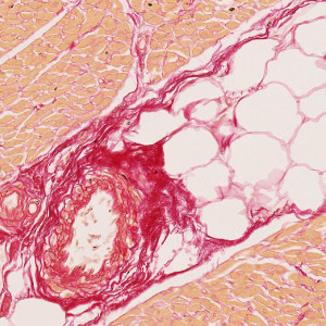
Sudden arrhythmic death syndrome (SADS) is a sudden, unexpected death in apparently healthy, young people where the underlying cause cannot be identified, see [1]. Sudden deaths are commonly attributed to cardiac abnormalities. Myocardial fibrosis, which includes an excess amount of collagen in the myocardial tissue, is a structural abnormality that may represent underlying cardiomyopathy causing sudden deaths, see [2]. Picrosirius red (PSR) is a stain that is commonly used to visualize collagen, see [3].
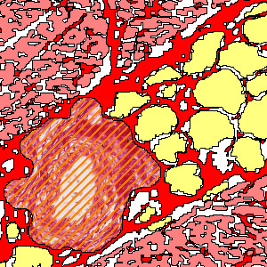
This APP is used for quantifying the amount of collagen, myocardial tissue, and fatty tissue in slides. It is also capable of detecting vessels in order to exclude them from the subsequent analysis so that they do not contribute to the outputs.
Details
Quantitative Output variables
The output variables obtained from this protocol are:
- Area of Collagen [μm2]
- Area of Myocardial Tissue [μm2]
- Area of Fatty Tissue [μm2]
- Total Tissue Area [μm2]
- Percentage of Collagen
- Percentage of Myocardial Tissue
- Percentage of Fatty Tissue
Workflow
Step 1: Load and run the APP “01 Tissue Detection” for detection of the tissue in the slide
Step 2: Load and run the APP “02 Large Vessels” for identification of larger vessels
Step 3: Load and run the APP “03 Small Vessels” for identification of smaller vessels
Step 4: Load and run the APP “04 PSR” for quantification of collagen, myocardial tissue, and fatty tissue
Methods
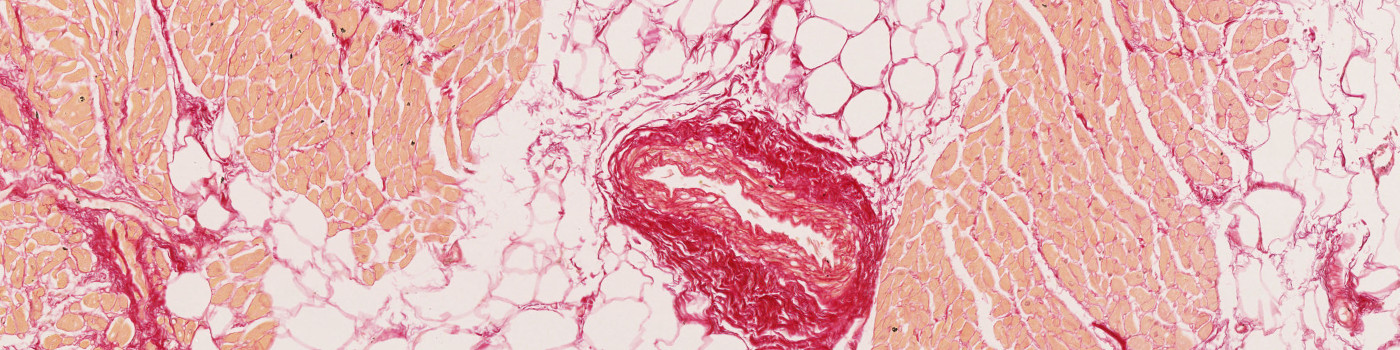
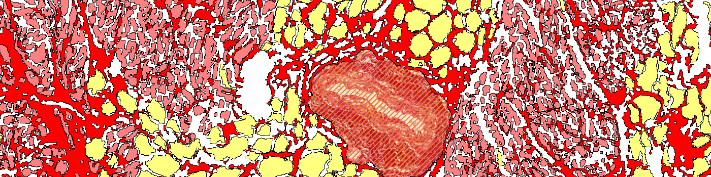
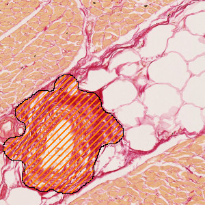
The APP consists of three protocols. The first two protocols detect large and small vessels, respectively, in order to exclude them from the subsequent analysis. The vessels are identified based on detection of lumens using the blue color band and by detection of surrounding layers of red PSR staining found using the contrast between the red and the green color bands. A lumen candidate is defined as an actual lumen if it is partly or fully surrounded by PSR staining. The vessel is set to be the lumen with associated red PSR staining. A morphological measure for circularity is used to remove false positives. The third protocol detects cardiac muscle tissue, collagen and fat cells. Cardiac muscle tissue is found using the contrast between the green and the blue color bands, collagen is found using a custom defined input band named fast red, and fatty tissue is found using the green color band. Different value intervals of these bands define the different classes of tissue. For fatty tissue, morphological operations are used to distinguish them from air wholes.
Staining Protocol
There is no staining protocol available.
Keywords
Picrosirius Red, PSR, Sudden Arrhytmic Death Syndrome, SADS, Cardiac, Digital Pathology, Image Analysis
References
USERS
This APP was developed for Dr. Chris Miles and Dr. Elijah Behr at St. George’s University of London
LITERATURE
1. Behr, E. R., et al. Sudden Arrhythmic Death Syndrome: A National Survey of Sudden Unexplained Cardiac Death. Heart 2007, 93 (5), 601–5, DOI.
2. Lecomte, D., et al. Isolated Myocardial Fibrosis as a Cause of Sudden Cardiac Death and Its Possible Relation to Myocarditis. J. Forensic Sci. 1993, 38 (3), 617-21.
3. Kiernan, J. A. Picrosirius Red Staining Protocol for Collagen. Accessed: June 12, 2019.