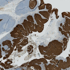
CK5 stained slide.
#10097
Phosphohistone H3 (PHH3) is a mitotic marker and has prognostic capabilities when applied to breast cancer tissue. The mitotic index can be obtained by PHH3-immunohistochemical staining, which can be used as a supplement for diagnosis. CK5 is overexpressed in tumors located in the oral cavity, oropharyngeal, hypopharyngeal and laryngeal areas [1] and can, thus, be used as a tumor marker.
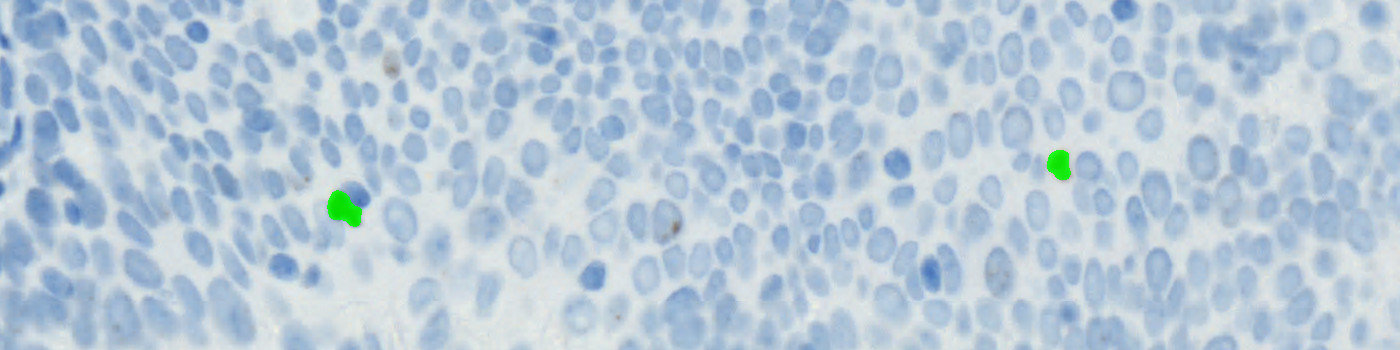
The “10097 – PHH3 + CK5 VDS, Neck Cancer” APP automatically detects and quantifies PHH3 positive nuclei within tumor regions that are identified based on the PHH3 and CK5 virtual double staining. Two serial sections stained for PHH3 and CK5, respectively, must be used in this APP.
CK5 stained slide.
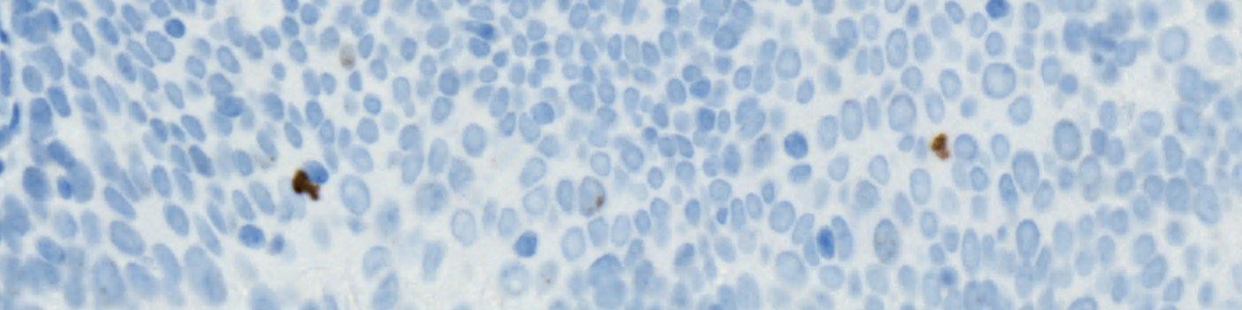
The serial PHH3 stained slide.

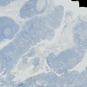
Illustration of the tissue alignment workflow linking the two differently stained serial sections together. This workflow requires the module Tissuealign™ with VirtualDoubleStaining™.
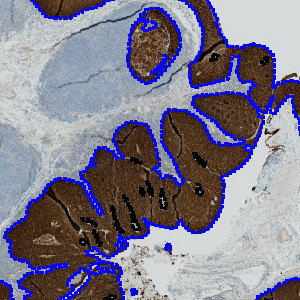
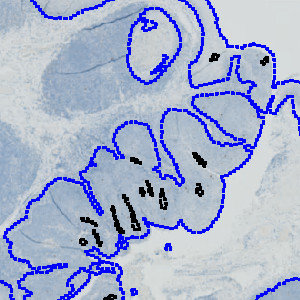
Tumor regions are automatically identified on the CK5 slide, and outlined with a blue ROI.
Auxiliary APPs
APP: “01 Tumor Detection”
The auxiliary APP ’01 Tumor Detection” is used for automatic tumor detection.
Quantitative Output variables
The output variables obtained from this protocol are:
Workflow
Step 1: Load and run the auxiliary APP for tumor region identification: “01 Tumor Detection”.
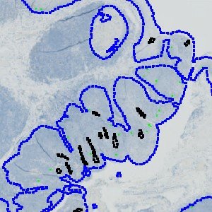
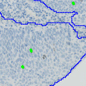
Step 2: Load and run the APP for identification of PHH3 positive nuclei: “02 Analyze”.
Methods
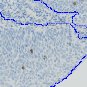
The PHH3 and CK5 serial sections are linked together and aligned via the Tissuealign™ workflow that allows for an easy user-assisted computational alignment. Tumor regions are then automatically identified based on the CK5 stained slide using the VirtualDoubleStaining™ technique. The tumor regions are overlaid on the PHH3 stained slide, and the subsequent analysis is now limited to the inside of the detected tumor regions only.
The PHH3 stained nuclei are detected based on the calculation of Hematoxylin and DAB color-deconvolution bands. These bands are used as the input parameters to a cell classification protocol identifying positively stained nuclei. Nuclei are managed with a postprocessing protocol to reduce the number of false positives and false negatives by merging closely connected nuclei and removing very small nuclei.
Staining Protocol
There is no staining protocol available.
Keywords
PHH3, Phosphohistone H3, CK5, IHC, VDS™, VirtualDoubleStaining™, neck cancer, digital pathology, image analysis.
References
LITERATURE
1. Woodcock-Mitchell, J., Eichener, R., Nelson, W. G., Sun, T. T. Immunolocalization of Keratin Polypeptides in Human Epidermis Using Monoclonal Antibodie, Journal of Cell Biology 1982, 95 (2), 580-588, DOI.