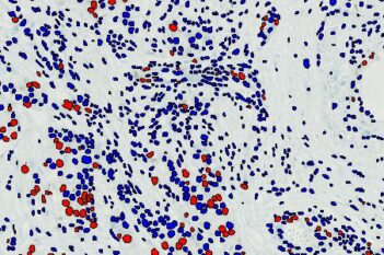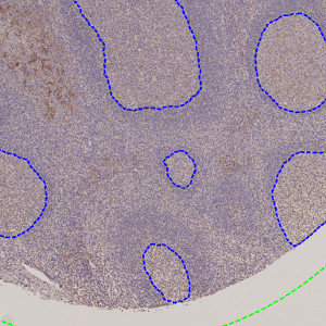
Outlined germinal centers (blue) on TMA core.
#10163
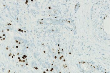
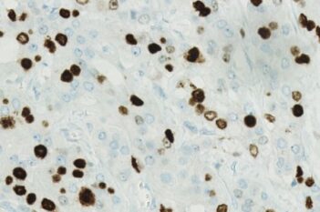
This Quickstart APP speeds analysis by automatically outlining germinal centers in tonsils. Trained on PD-L1 (membrane) and Ki-67 (nuclear) staining, this APP is generalizable to other stains. As with all of our Quickstart APPs, they are fully customizable and allow for scalable image analysis with the batch analysis feature.
Working with multimodal data sets or TMAs? No problem. Run these APPs after using Tissuealign™ feature to align your images at the cellular level, or the Tissuearray™ feature to automate de-arraying of your tissue cores.
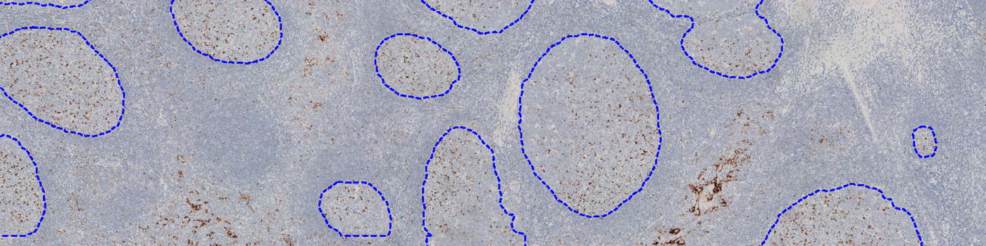
Outlined germinal centers (blue) on TMA core.
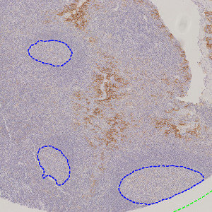
Outlined germinal centers (blue) on TMA core.
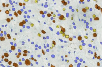
Quantitative Output variables
Germinal Center Area [mm²]
Workflow
After loading your image, perform the following steps:
Step 1: Open the “10163 IHC, Germinal Cancer Detection.”
Step 2: Click “Run APP” or use the batch analysis feature to queue the APP and continue working on other images