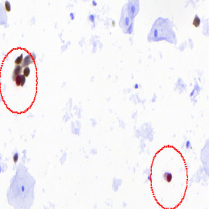
Raw image.
#10028
Human Papillomavirus (HPV) is the main cause of cervical cancer and its precursor lesions. Tests for HPV are being widely used in adjunctive testing in cervical cytology specimens with atypical cells of undetermined significance (ASCUS) and low grade of intraepithelial lesion (LSIL) in order to identify the women at true risk of harbouring histological high grade lesions, who require treatment.
It is well known that tests for HPV are highly sensitive but lack specificity, especially in women with LSIL. Immunocytochemical dual staining for p16 and Ki-67 is a promising test, and the few studies published so far have showed high specificity and retained sensitivity for identifying high grade lesions.
However, the evaluation by microscope is subjective and labour-intensive, and preliminary results have shown that the test suffers from limited reproducibility. With the “p16+Ki-67, Cervical Cancer, ICC” APP automatic detection of the p16/Ki-67 dual stained cells is possible thereby converting manual inspection of all cells on a slide to a final review of suspicious cells selected by the computer.
The APP can be used for quantifying the p16/Ki-67 dual stained cells and outputs a score for each p16/Ki-67 nuclei based on the degree of positively stained cytoplasm.
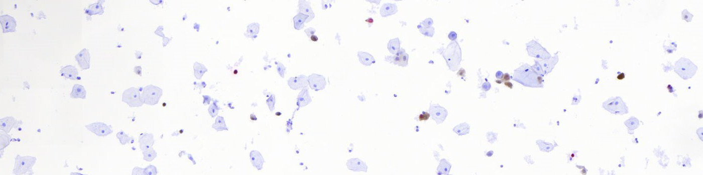
Raw image.
Result of analysis with the auxiliary ROI identification APP. Only p16/Ki-67 dual stained cells are marked as relevant ROIs for further analysis.
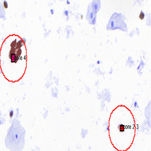
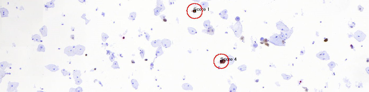
Result of analysis with the APP: Each p16/Ki-67 dual stained nuclei has been given a score from 0-4 based on the cytoplasmic staining. Notice how only cells within the detected ROI are analyzed providing faster analysis.
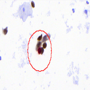
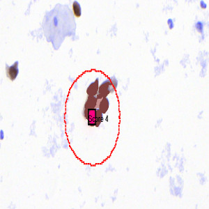
Zoom in on one particular p16/Ki-67 dual stained cell.
Auxiliary APPs
ROI DETECTION
APP: “01 Cervix 5X detect”
This APP is used for automatically creating a ROI around the positively stained nuclei and their cytoplasm.
The APP identifies regions of interests (ROIs) based on the p16 (DAB), Ki-67 (Fast Red) and Hematoxylin color-deconvolution bands. The color-deconvolution bands are used as input parameters to a naïve Bayesian classifier that identifies positively stained nuclei and their cytoplasm as well as negative cells. A ROI is then automatically created around the positively stained cells only, so that further processing of the image is limited to be within this area making the processing time considerably faster (see FIGURE 1 and 2).
REMOVE ANNOTATIONS
APP: “03 Remove Annotations”
This APP can be used for automatic and fast removal of the scoring annotations if desired.
Quantitative Output variables
The output variables obtained from this protocol include:
Workflow
Step 1: Load the APP for ROI identification: “01 Cervix 5X detect”.
Step 2: Load the APP for identification and scoring of p16/Ki-67 dual stained cells: “02 Cervix Analyze”.
Step 3: If desired, load the auxiliary APP for removal of scoring annotations: “03 Remove Annotations”
Methods
The APP detects and scores the dual stained p16/Ki-67 cells. The detection and scoring of dual stained p16/Ki67 objects is based on the calculation of p16 (DAB), Ki-67 (Fast Red) and Hematoxylin color-deconvolution bands. These bands are used as the input parameters to a naive Bayesian probabilistic classifier identifying positively stained nuclei, and unsurely or positively stained cytoplasm. Objects are defined by connected regions of red pixels, restricted by the 4-pixel connectivity rule, with a profile area corresponding to a typical relevant nucleus. In the protocol, the scoring of the detected object nucleus is solely based on the characteristics of the cytoplasm that surrounds the detected red nucleus, in the following way (see FIGURE 3):
– 0 (red nucleus without positive or unsure cytoplasm),
– 1 (red nucleus with unsure cytoplasm),
– 2-3 (red nucleus with positive and unsure cytoplasm) and
– 4 (red nucleus with positive cytoplasm).
Detected nuclei and cytoplasm, of either of the scored detections, in close conjunction are defined as larger clusters and handled separately.
Staining Protocol
Immunocytochemical simultaneous double staining for the cell cycle regulator p16INK4a and the active cell proliferation marker Ki67 was performed using the CINtec PLUS kit (Roche, Germany).
Keywords
Ki-67, p16, dual stained cells, HPV, cervix, digital pathology, image analysis
References
USERS
This APP was developed in cooperation with Marianne Waldstrøm and Christina Braad Petersen, Department of Clinical Pathology, Vejle Hospital.
LITTERATURE
There are no references.