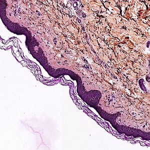
Orcein stained skin section.
#10020
Epidermal thickness, and the content of collagen/elastin in the dermal layer, and the relationship to age, gender, skin type, pigmentation, blood content, smoking habits and body site is important in dermatologic research. The epidermal thickness is often affected during skin diseases, such as psoriasis, and its measurement can therefore be a relevant parameter in the assessment of disease progression and treatment effect.
The thickness of the epidermis can vary substantially even within a tissue section. For this reason, the arithmetic mean thickness is estimated as the epidermis area divided by the length of the epidermis.
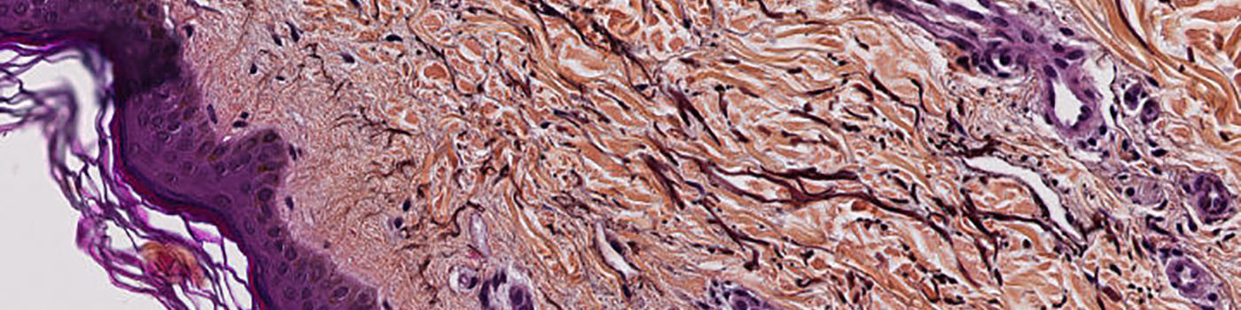
Orcein stained skin section.
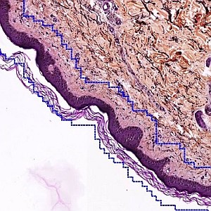
Orcein stained skin section, with rough outlining of the epidermis. This is performed automatically.
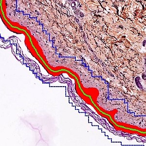
The area (red) and length (green) of the epidermis is used for calculating its arithmetic mean thickness.
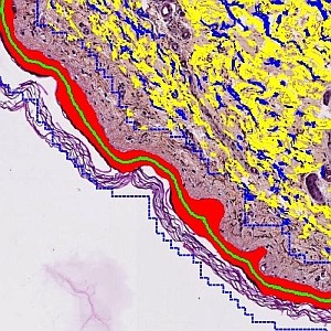
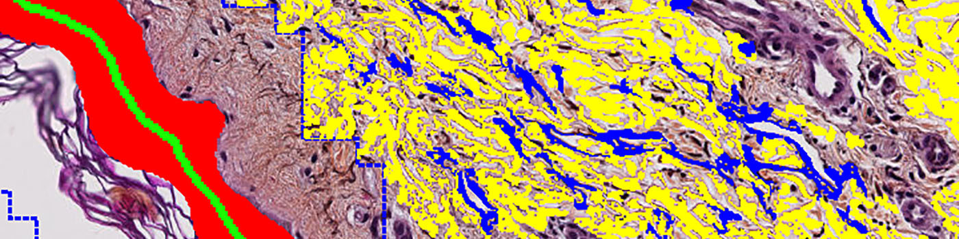
Additional detection of the elastin (blue) and collagen (yellow) in the skin section. This is only performed in the dermis.
Auxiliary APPs
Auxiliary APPs are used for additional process steps, e.g. finding Region of Interest (ROI).
TISSUE DETECT
The auxiliary APP ’01 Tissue ROI’ can be used to automatically detect the region of interest (ROI). This is to save time in the subsequent analysis steps. The analysis is carried out at a lower magnification and finds larger pieces of tissue. Dust and small artifacts are ignored. There are no quantitative output variables from this step (see FIGURE 1 and 2).
ELASTIN/COLLAGEN QUANTIFICATION
The auxiliary APP ’03 Analyze Collagen Elastin’ segments the detected dermis into different components: collagen and elastin.
Quantitative Output variables
The endpoint for this analysis is the arithmetic mean thickness of the epidermis:
The analysis could be extended to also include area ratio (epidermis to total tissue ratio) or other relevant measures.
Workflow
Step 1: Load auxiliary APP for tissue detection ’01 Tissue ROI’ and run on the image to process. This outlines the tissue with a Region Of Interest (ROI) and saves time in the subsequent analysis steps.
Step 2: Load quantification APP ’02 Analyze’ and run on the image to detect the epidermis and calculate its thickness.
Step 3 (optional): Load quantification APP ’03 Analyze Collagen Elastin’ and run on the images to segment the dermis into collagen and elastin.
Methods
Prior to the image analysis, it is important to outline the region of interest, thereby excluding most of the skin, which is not epidermis from the quantitative analysis. This outlining is performed automatically, but could also be done manually. It does not need to be extremely precise, as long as it eliminates tissue areas of the same color as the epidermis. The segmented epidermal layer is skeletonized, thereby generating its midline (see FIGURE 3). The length of the interface between the skeleton and the original epidermis corresponds to two times the length of the epidermis. Finally, a segmentation of the outlined dermis is performed to detect the elastin and collagen content.
Keywords
Skin, Epidermis, Thickness, Psoriasis, quantitative, digital pathology, image analysis, elastin, collagen.