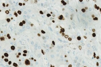
Breast tissue before processing with this APP.
#10188
This Quickstart APP offers a fast and accurate way to segment nuclei and measure 3,3′-Diaminobenzidin (DAB) intensity in tissue samples, giving you a reliable starting point for biomarker quantification. Designed for high-resolution analysis, it delivers single-cell outputs that can be reviewed with the interactive plotting tool for quality control, making it ideal for studies involving ER, PR, Ki-67, and other nuclear markers.
It can be run in a sequence, such as after performing tissue detection (see “Related APPs” below). As with all of our Quickstart APPs, your image analysis can be scaled with the “batch analysis” feature available on both Discovery and Phenoplex™.
Working with multimodal data sets or TMAs? No problem. Run this APP after using the Tissuealign™ feature to align your images at the cellular level, or the Tissuearray™ feature to de-array your tissue cores.
Breast tissue before processing with this APP.
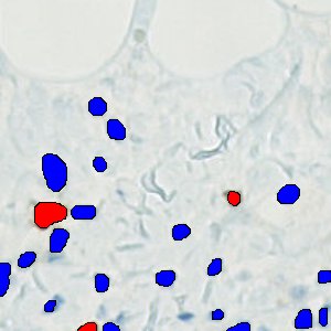
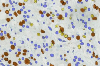
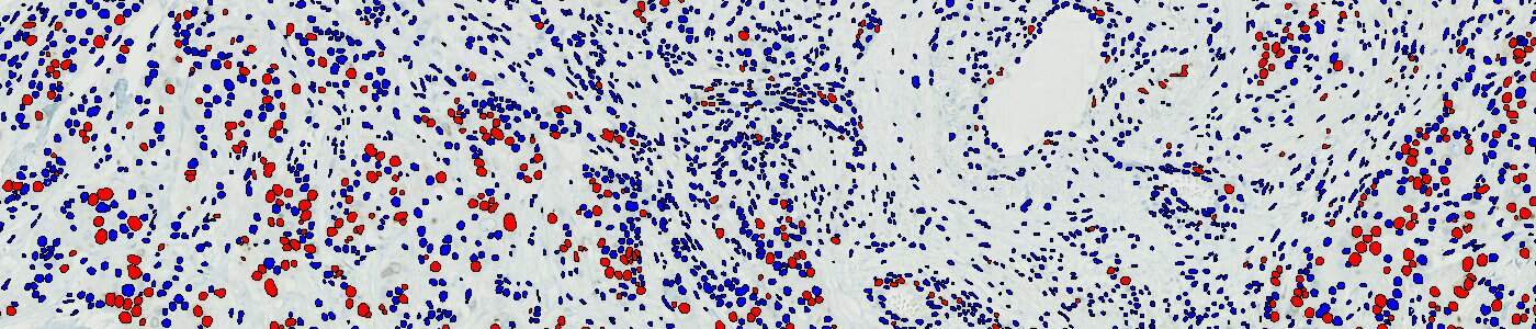
Post processed. Blue cells are DAB – and red cells are DAB +.
Quantitative Output variables
Total nuclei count, positive percentage, and per nuclei DAB measurement.
Workflow