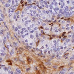
Close up mouse breast tissue displaying negative nuclei (blue) and PEG positive areas (brown).
#10084
In magnetic resonance imaging (MRI) a contrast agent is used to enhance soft tissue of interest. In a study, see [1], a method was developed to detect a nanoparticle based contrast agent, specifically designed to target tumor tissue. Polyethylene glycol (PEG) coats the nanoparticles and can be targeted by antibodies through immunohistochemistry (IHC) staining, using diaminobenzidine (DAB).
This APP automatically detects the PEG positive areas in the tissue as well as the ratio compared to the total tissue area, which should give an indication of the tumor growth.
Close up mouse breast tissue displaying negative nuclei (blue) and PEG positive areas (brown).
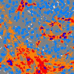
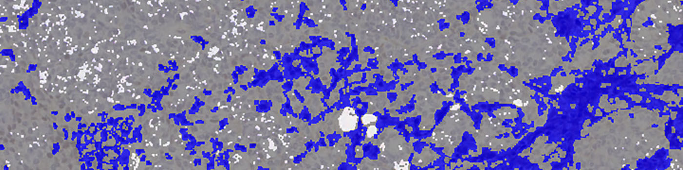
The original image was classified into tissue or PEG positive areas with high, medium and low DAB chromogen intensity.
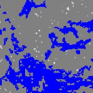
A post processing step was used to change the low, medium and high PEG intensities into one, which was used in the calculation step.
Quantitative Output variables
Methods
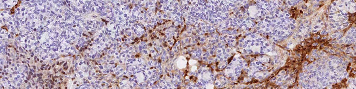
Hematoxylin was used to stain nuclei (blue) and the brown chromogen diaminobenzidine (DAB) was used to visualize PEG-positive tissue areas as described in the staining protocol (see FIGURE 1 and 2). VISIOmorph was used to calculate the amount of PEG in the tissue expressed as % of tissue area. The preprocessing consisted of DAB and Hematoxylin color de-convolution. The blue color channel was used to detect the remaining tissue. Three levels of DAB chromogen intensity (high, medium and low) was classified using the Bayesian classification method (see FIGURE 2). This was done to allow for the possibility to fine tune the configuration and/or to analyze the PEG intensities separately. However, no significant differences were observed when using separate PEG intensities when comparing tumor tissue. Therefore, in the final settings the PEG intensities were added together and the total amount of PEG in the tissue was calculated (see FIGURE 3).
Staining Protocol
As described in Eriksson et al, see [1]:
Paraformaldehyde fixed muscle and tumor tissues collected before injection and at 30 minutes and 2-4 h post-dose were paraffin embedded and thereafter cut (4 µm) and positioned on glass tissue slides. Prior to staining, the slides were placed in an antigen-retrieval solution using an automated pre-treatment module (PT-Link; Dako, Glostrup, Denmark). Slides were stained for PEG in an automated immunohistochemistry robot (Autostainer; Dako) using a primary rabbit anti-PEG-B-47 antibody (Abcam, Cambride, MA) at a concentration of 0.6 µg/ml and a secondary goat-anti rabbit antibody (Dako) at 1:200 dilution. Slides were counterstained with hematoxylin, dehydrated and mounted. The stained slides were digitalized using an ultra-high-resolution slide scanner (Aperio Scan Scope, Aperio Technologies, CA USA) and total PEG-staining was quantified with the VisomorphDP software (Visiopharm, Hoersholm, Denmark) and total PEG-staining was quantified with the Visomorph software (Visiopharm, Hoersholm, Denmark).
Additional information
This APP was developed by Caroline Sandén at Medetect AB in collaboration with Dept. Airway Inflammation at Lund University, for Spago Nanomedical AB.
Spago Nanomedical is developing a nanoparticle based contrast agent, SpagoPix, for diagnosis of tumors with MRI. The proprietary nanoparticles provide exceptional MR signal enhancement and selectively target tumor tissue via the passive EPR effect (Enhanced Permeability and Retention), which results in improved tumor detection over current contrast agents. The tumor targeting properties were illustrated in a study published in PlosOne (see references) where MRI was used to locate tumors in a clinically relevant mouse breast cancer model (MMTV-PyMT) injected with SpagoPix. The imaging results were supplemented and confirmed with quantification of SpagoPix by chemical analysis (ICP) and with immunohistochemistry using antibodies directed against the polyethylene glycol (PEG) surface coating on the nanoparticles.
Keywords
Polyethylene glycol, nanoparticles, immunohistochemistry
References
LITERATURE
1. Eriksson, P-O., et. al., Novel Nano-Sized MR Contrast Agent Mediates Strong Tumor Contrast Enhancement in an Oncogene-Driven Breast Cancer Model, Plos One 2014, 9 (10), e107762, DOI