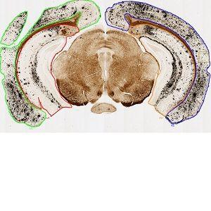
Low magnification overview image showing the 4 regions of interest from a coronal brain section: Left cerebral cortex (green), right cerebral cortex (blue), left hippocampus (red), and right hippocampus (yellow).
#10032
Alzheimer disease (AD) is the most common form of dementia, manifesting with a wide range of symptoms. This progressive disorder is incurable and as it worsens it will lead to cognitive decline, loss of body function and ultimately death.
The exact mechanisms of AD are not well understood, but it is known that accumulation of the amyloid-β (Aβ) peptide plays an essential role in the pathogenesis.
There is currently no cure for AD, and treatment will only relieve the symptoms of the disease. Research focused on anti-Aβ immunotherapies in animal models indicates that both active and passive immunization against Aβ will improve cognitive function and clear the parenchymal accumulation of amyloid (plaques) in the brain.
Although immunization against Aβ holds promise as a disease-modifying therapy for AD, it is associated with an undesirable accumulation of amyloid in the cerebrovasculature (i.e., cerebral amyloid angiopathy, CAA) as well as a heightened risk of micro-hemorrhages. Given the likelihood that immunotherapy-solubilized amyloid redistributes from the parenchymal plaques to the cerebral vasculature, it is possible that the amyloid clearance from prolonged intracerebroventricular (icv) infusions of low-dose anti-Aβ antibodies is sufficient to gradually prevent a substantial accumulation in the cerebral vasculature, and hence will not result in CAA, see [1].
The clearance of amyloid plaque as well as the degree of CAA is examined with this APP.
The Campbell-Switzer stain was used to visualize amyloid plaques in the parenchyma and CAA. Positive staining for plaques was verified using immunohistochemical detection of Aβ.
With this method, plaques will appear in black while CAA appears in a reddish brown.
Low magnification overview image showing the 4 regions of interest from a coronal brain section: Left cerebral cortex (green), right cerebral cortex (blue), left hippocampus (red), and right hippocampus (yellow).
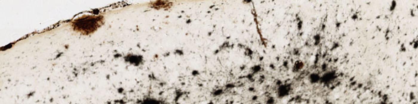
10x field-of-view scaled to fit this space, showing the image before classification.
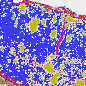
10x field-of-view scaled to fit this space, showing the initial classification based on the Bayesian classifier.
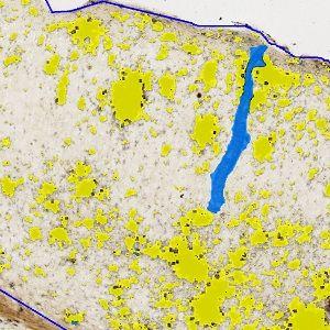
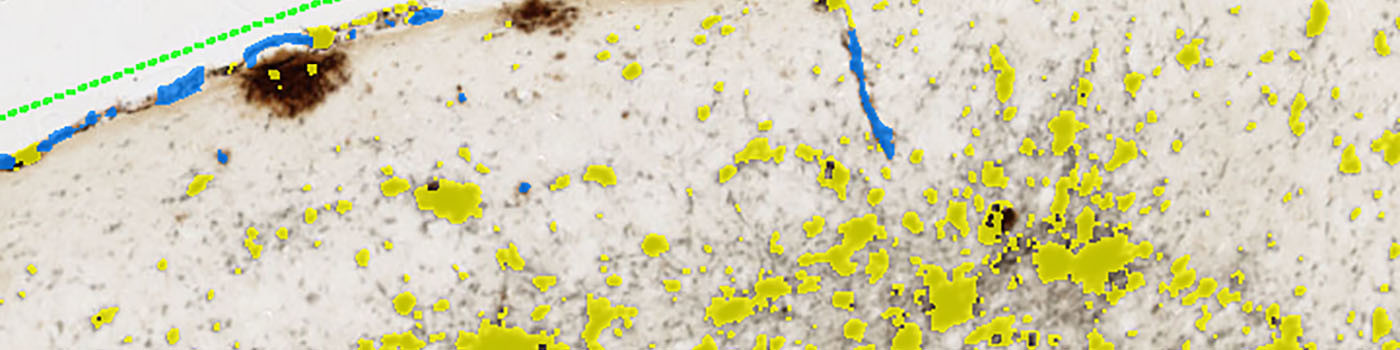
10x field-of-view scaled to fit this space, showing the result after post processing. The identified CAA is overlayed with blue, compared to amyloid plaques that are overlaid with yellow.
Quantitative Output variables
The output variables obtained from this protocol include 3 calculations for each of the 4 ROI’s Left Cortex (LC), Right Cortex (RC), Left Hippocampus (LH) and Right Hippocampus (RH):
Methods
The sections analyzed are coronal brain sections. Before applying the image analysis protocol, the Regions of interest (ROIs), the left and right of Cerebral Cortex and Hippocampus, must be outlined manually as shown in FIGURE 1.
The protocol uses a Bayesian classifier to distinguish between positive amyloid stain (black), CAA (brown), and background. Post-process steps will remove all unnecessary labels as well as recognize CAA areas based on shape, surroundings, and size.
By analysis of the stained brain sections, the aim is to quantify the total relative stain area i.e., the combined area of parenchymal plaques and CAA (in %) relative to the cerebral cortex or hippocampus area, as well as the CAA relative stain area i.e., the area of CAA (in %) relative to the cerebral cortex or hippocampus area.
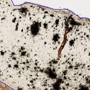
FIGURES 2 and 3 show a field of view before classification, and after classification before post processing, respectively. An area identified as CAA can be seen after post processing in FIGURE 4.
Keywords
Alzheimer Disease, Amyloid plaque, Aβ, immunization, Campbell-Switzer, plaque clearance, CAA, quantitative, digital pathology, image analysis.
References
LITERATURE
1. Thakker, D.R., et. al., Intracerebroventricular amyloid-β antibodies reduce cerebral amyloid angiopathy and associated micro-hemorrhages in aged Tg2576 mice, PNAS 2009, 106 (11), 4501-6, DOI