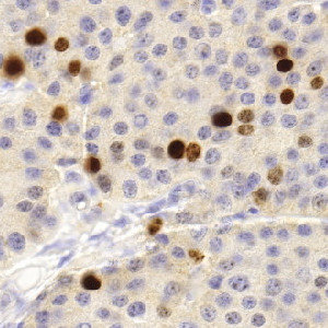
Original image of Ki-67 stained neuroendocrine neoplasm.
#10143
The Ki-67 protein is associated with cellular proliferation, and the protein is present in the nucleus of all cells that are in the active phase of the cell cycle, but absent in resting cells, see [1]. The cell proliferation rate can be assessed by Ki-67-immunohistochemical (IHC) staining, and this can be correlated to the tumor grade and the clinical course. Ki-67 proliferative index is suggested to be an important prognostic variable and is used as a grading parameter for neuroendocrine neoplasms, see [2].
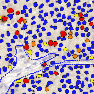
The APP detects Ki-67 negative and positive nuclei, classifies the positive nuclei into 1+, 2+ and 3+ categories, and calculates the proliferation index for neuroendocrine tissue stained for Ki-67.
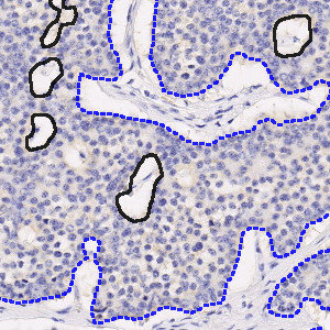
Original image of Ki-67 stained neuroendocrine neoplasm.
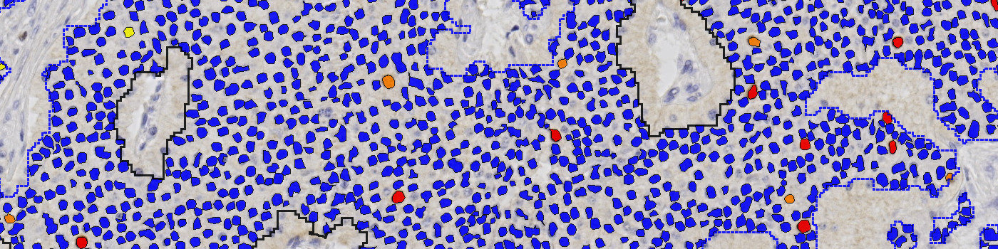
Analyzed image of Ki-67 stained neuroendocrine neoplasm with automatic tumor outline.
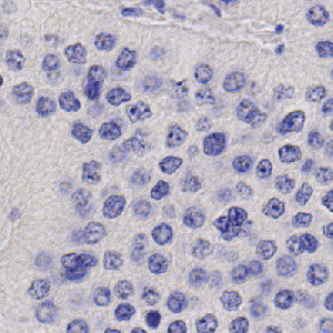
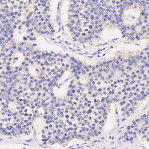
Original image of Ki-67 negative neuroendocrine neoplasm.
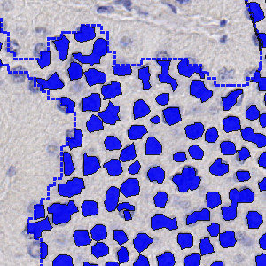
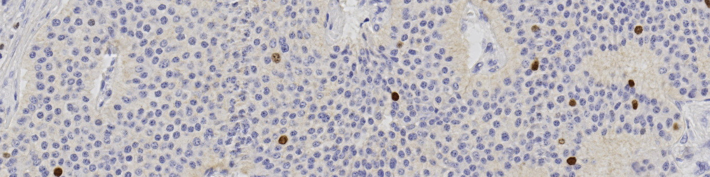
Analyzed image of Ki-67 negative neuroendocrine neoplasm, negative nuclei shown in blue.
Quantitative Output variables
The output variables obtained from this protocol include:
Workflow
Step 1: Load and run the APP “01 Tumor Detection” to outline tumor ROIs and adjust manually as needed.
Step 2: Load and run the APP “02 Analyze” to analyze nuclei in tumor region(s).
Methods
The APP works within an automatically or manually outlined tumor region of interest (ROI). The first image processing step involves a segmentation of all nuclei in the ROI. This is done by using an HDAB-DAB color deconvolution band to detect positively stained nuclei and a multiplication of the red and blue color band to detect negative nuclei. A method for nucleus separation which is based on shape, size and nuclei probability is used, employing a fully automated watershed-based nucleus segmentation technique. As a post-processing step, nuclei areas that are too small are removed. The image obtained after post-processing is used to determine the output variables.
Staining Protocol
There is no staining protocol available.
Keywords
Ki-67, Ki67, proliferation rate, immunohistochemistry, quantitative, digital pathology, image analysis, neuroendocrine, NET
References
USERS
The APP was developed for Ph.D Mohamed Ibrahim, University of Gothenburg.
LITERATURE
1. T.Scholzen et al. The Ki-67 protein: from the known and the unknown, J. Cell Physiol 2000, 182 (3), 311-22, DOI.
2. Nadler, A., Cukier, M., Rowsell, C., et al. Ki-67 is a reliable pathological grading marker for neuroendocrine tumors, Virchows Archiv 2013, 462 (5), 501-5, DOI.