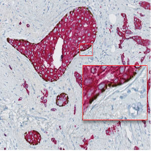
Image of a slide physically double stained with p63 (brown chromogen) and CK7/19 (red chromogen). Notice how only some structures display p63 positive nuclei.
#10101
Whether a cancer is invasive or non-invasive will determine the treatment choices and how a patient might respond to the treatment he or she receives. Most breast cancers are invasive, but in some cases both invasive and non-invasive cancer can be seen in the same specimen, see [1]. In these cases it is necessary with a tool that can distinguish between the invasive tumor components and the non-invasive tumor components, such as ductal carcinoma in situ (DCIS), so that the biomarker expression, e.g. Ki-67 or ER, within the invasive cancer, can be accurately assessed.
The “Invasive Tumor Detection (PDS)” APP provide means for distinguishing between invasive tumor and non-invasive tumor based on a slide physically double stained (PDS) with the myoepithelial cell nuclear marker p63 in brown and a cytokeratin (CK) tumor marker in red, e.g. CK7/19. With VirtualDoubleStaining™ this APP can be combined with other APPs intended for analysis of biomarker expression within invasive tumor components.
Image of a slide physically double stained with p63 (brown chromogen) and CK7/19 (red chromogen). Notice how only some structures display p63 positive nuclei.
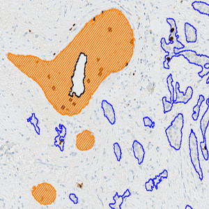
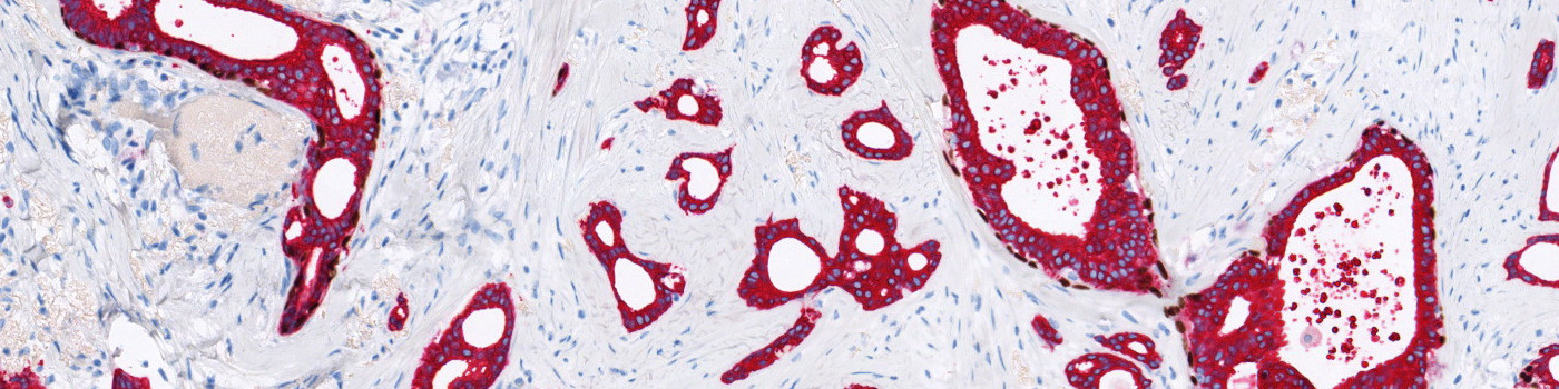
Automatic identification of the tumor components based on the red CK7/19 staining. Identified tumor is marked with a blue region of interest (ROi).
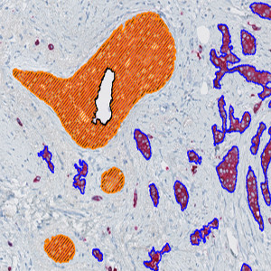
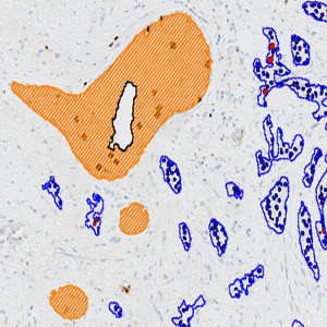
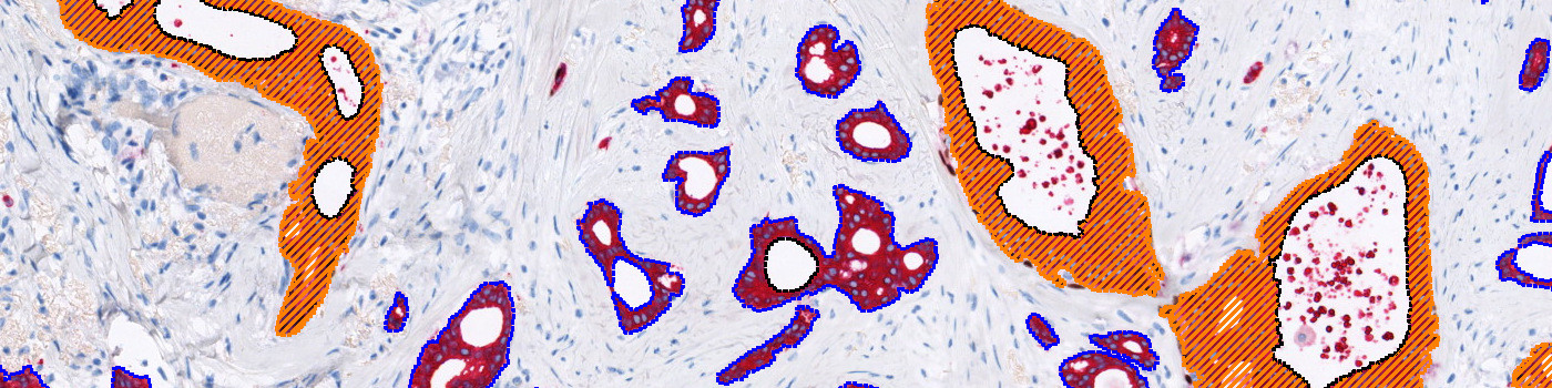
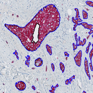
Automatic separation of tumor components into invasive and non-invasive tumor components (e.g. ductal carcinoma in situ) based on the joint p63 and CK7/19 staining. The invasive tumor components are marked with the blue ROI, while the non-invasive tumor components are marked with an orange ROI.
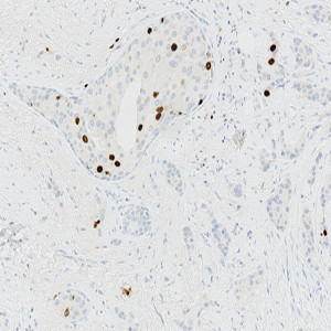
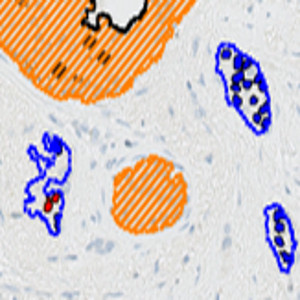
A slide stained with Ki-67 and serial to the p63+CK7/19 slide showed in FIGURE 1.
Auxiliary APPs
Protocol: “01 Tumor Detection”
The protocol “01 Tumor Detection” is used for automatic tumor detection.
Protocol: “02 Invasive tumor detection”
The protocol “02 Invasive Tumor Detection” is used for distinguishing the tumor components into invasive and non-invasive tumor components.
Protocol: “03 Invasive Tumor Merge”
The protocol “03 Invasive Tumor Merge” is used for merging small invasive tumor regions with non-invasive tumor regions if regions are split due to tiling
Quantitative Output variables
The output variables obtained from this APP are:
Workflow
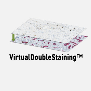
Without VirtualDoubleStaining™:
Step 1: Load and run the protocol “01 Tumor Detection” for tumor identification
Step 2: Load and run the protocol “02 Invasive Tumor Detection” for detection of invasive tumor components.
Step 3: Load and run the protocol “03 Invasive Tumor Merge”
With VirtualDoubleStaining™:
Step 1: Align the p63+CK section with a serial section stained with a desired marker
Step 2: Load and run the protocol “01 Tumor Detection” for tumor identification
Step 3: Load and run the protocol “02 Invasive Tumor Detection” for detection of invasive tumor components
Step 4: Load and run the protocol “03 Invasive Tumor Merge”
Step 5: Load and run the protocol suited for analysis of the slide stained with a desired marker within the invasive tumor components, e.g. APP “10004 – Ki-67, Breast Cancer”
Methods
The APP consists of three protocols. The first protocol detects all tumor present on the p63+CK slide using a linear Bayesian classification that identifies everything stained red as tumor (see FIGURE 2). The second protocol refines this tumor identification by discriminating between tumor components that are positive for both CK and p63, and tumor components that are only positive for CK. The tumor components only positive for CK are identified as the invasive tumor components (see FIGURE 3). The third protocol merges small invasive regions with non-invasive regions if regions are split due to tiling.
If a slide serial to the p63+CK slide and stained with e.g. Ki-67 or any other marker is available, it is possible, using VirtualDoubleStaining™, to conduct an analysis where the information present on each of the slides is used in a combined way. For example, in the case of having a Ki-67 slide that is serial to the p63+CK slide, it would be possible to identify the invasive and non-invasive tumor components on the Ki-67 slide based on the information present on the p63+CK slide. With this method it would then be possible to do a Ki-67 quantification within the invasive tumor components only (see FIGURE 4-8).
Staining Protocol
The slides showed here have been stained with p63 (clone 4A4, brown chromogen) from Ventana, CK7 (clone OV-TL12/30, red chromogen) from Dako, CK19 (clone A53-B/A2-26, red chromogen) from AH-Diagnostics, and Ki-67 (clone 30-9) from Ventana.
Keywords
Invasive tumor detection, Ductal carcinoma in situ, Physical double staining, VirtualDoubleStaining™, VDS™, Ki-67, IHC
References
LITERATURE
1. Breastcancer.org. Non-Invasive or Invasive Breast Cancer. (Last modified: 19. September 2018).