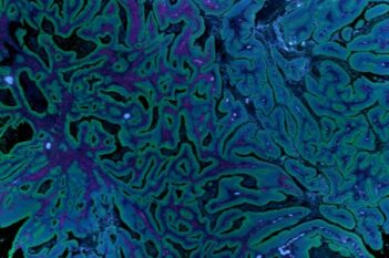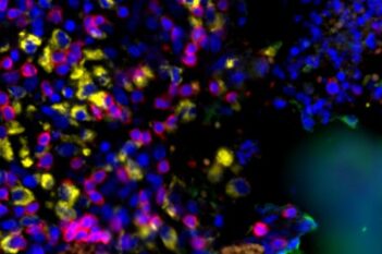
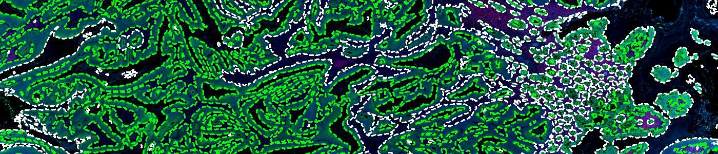
Multiplex GeoMx® DSP image before running one-click panCK detection.
#10194
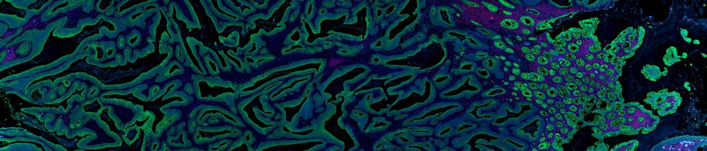
This Quickstart APP detects and outlines epithelial and the remaining tissue, including stroma regions, in images from Bruker’s GeoMx® DSP using the pan cytokeratin (panCK) channel. Tailor downstream analysis on specific tissue compartments or distinguish between cell populations based on their spatial context.
As with all of our Quickstart APPs, your image analysis can be scaled by queueing APPs to run in the background on an unlimited number of images—this is possible with the “batch analysis” feature available on both Discovery and Phenoplex™.
Working with multimodal data sets or TMAs? No problem. Run this APP after using the Tissuealign™ feature to align your images at the cellular level, or the Tissuearray™ feature to de-array your tissue cores.
After analysis is complete, you may explore your results with the Phenoplex™ Guided Workflow or the Data Exploration and QC tool (available on Discovery and Phenoplex™).
Multiplex GeoMx® DSP image before running one-click panCK detection.
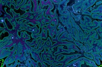
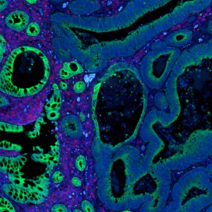
The tissue sections outlined in white correspond to sections positive for panCK.
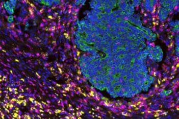
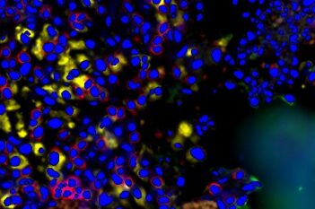
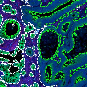
Another section of the same image before running this APP.

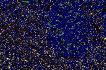
This APP accurately shows tissue not staining for pan cytokeratin (green) and tissue sections positive for the marker.
Quantitative Output variables
Workflow