
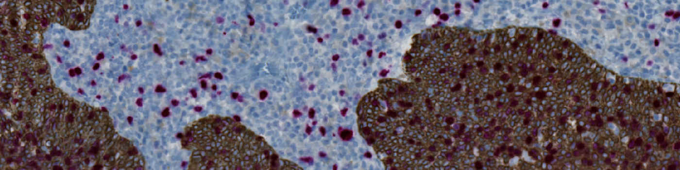
Raw image. Upper left side shows normal neck tissue. Lower right shows tumor area.
#10094
The Ki-67 protein is associated with cellular proliferation, and the protein is present in the nucleus of all cells that are in the active phase of the cell cycle, but absent in resting cells, see [1]. The cell proliferation rate can be assessed by Ki-67-immunohistochemical (IHC) staining, and this can be correlated to the tumor grade and the clinical course for the patient, see [2]. To identify the Ki-67 positive cells within the tumor regions on an image, these regions must be outlined manually, which can be a tedious and time-consuming task. However, tumor detection can be automated by performing a physical double staining (PDS) with cytokeratin 5 (CK5). CK5 is overexpressed in tumors located in the oral cavity, oropharyngeal, hypopharyngeal and laryngeal areas, see [3]. CK5 can be used as a tumor marker, and Ki-67 nuclei can thus be classified as belonging to a tumor based on the staining of the surrounding tissue. By using PDS with CK5, automated segmentation of relevant nuclei may be more precise and reproducible, see [4].
This protocol automatically detects and quantifies Ki-67 positive nuclei within neck tissue tumor regions. No manual outlining of the tumor is needed, since the tumor region is automatically identified based on the CK5+Ki-67 PDS. The protocol provides the number of positive nuclei within the tumor region as well as the ratio compared to the total number of nuclei within the tumor region.
Raw image. Upper left side shows normal neck tissue. Lower right shows tumor area.
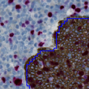
Result of analysis with the Auxiliary ROI detect APP. The tumor area has clearly been outlined by a blue dashed ROI.
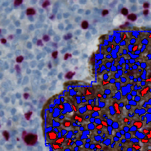
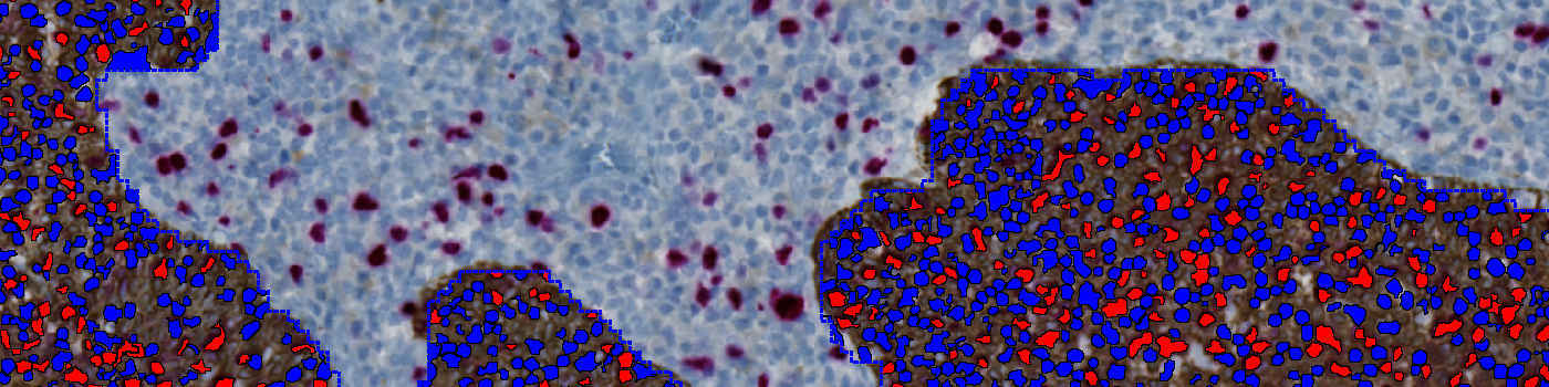
Result of analysis with the APP. Only areas within the detected tumor ROIs are analyzed resulting in a faster analysis. The Ki-67 positive nuclei are labeled with a red colored label and the negative nuclei with a blue colored label.
Auxiliary APPs
ROI detection:
APP: “01 ROI detect”
The auxiliary APP ’01 ROI detect’ is used to automatically outline the CK5 stained tumor regions as the regions of interest for the subsequent analysis.
Quantitative Output variables
The output variables obtained from this protocol are:
Workflow
Step 1: Load the auxiliary APP for ROI identification (“01 ROI detect”)
Step 2: Load the APP for identification and scoring of Ki-67 stained nuclei (“02 Analyze”)
Methods
The first image processing step involves an automated detection/outlining of tumor area, i.e. the region of interest (ROI) (see FIGURE 2). The nuclei are subsequently detected inside the ROI as either Ki-67 positively stained nuclei or negatively stained nuclei (see FIGURE 3). The total number of cells and the positive nuclei ratio is calculated and provides a proliferation index from 0-100% indicating the number of cells in a tumor that are dividing.
The APP detects and scores the dual stained Ki-67 cells. The detection and scoring of dual stained Ki67 objects is based on the calculation of p16, Ki-67 and Hematoxylin color-deconvolution bands. These bands are used as the input parameters to a threshold classifier identifying positively or negatively stained nuclei. Nuclei are managed with a postprocessing protocol to reduce the number of false positives and false negatives by merging closely connected nuclei and remove very small nuclei (see FIGURE 3).
Staining Protocol
There is no staining protocol available.
Keywords
Ki-67, CK5, dual stained cells, physical double staining, neck cancer, digital pathology, image analysis
References
LITERATURE
1. Sholzen, T., Gerdes, J. The Ki-67 protein: from the known and the unknown, Journal of Cellular Physiology 2000, 182 (3), 311-322, DOI.
2. Nordic Immunohistochemical Quality Control (NordiQC), Ki67. (Last updated: 02.07.09).
3. Woodcosk-Mitchell, J., Eichner, R., Nelson, W. G., Sun, T. T. Immunolocalization of Keratin Polypeptides in Human Epidermis Using Monoclonal Antibodies, Journal of Cell Biology 1982, 95 (2), 580-588, DOI.
4. Bentzer, N. K., et. al. Ki67-labeling index in breast cancer – An immunohistochemical pilot study using double staining to evaluate proliferation rate, APMIS 2010, 118, 15-16, DOI