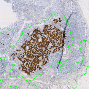
PCK positive regions are roughly outlined for further analysis (shown in green).
#10153
Developed for metastasis detection in cytokeratin stained lymph nodes
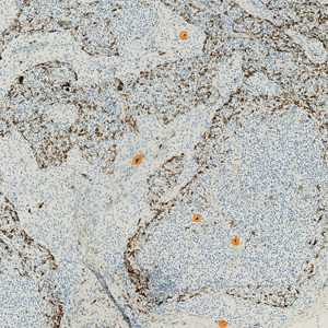
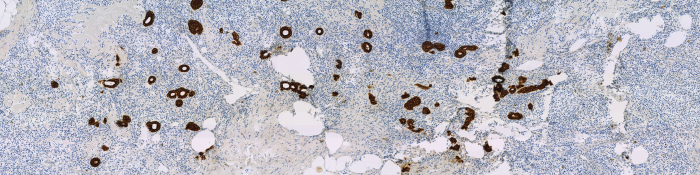
Tumor staging, diagnosis, and treatment choices depend, for most cancer types, on the size of the primary tumor and whether it has metastasized to regional lymph nodes. Lymph node metastasis can be identified using a pancytokeratin (PCK) stain as a cytoplasmatic marker, coloring them brown.
The APP identifies cytokeratin positive cells and calculates the diameter or area, of the largest tumor. Images can easily be sorted based on their size, e.g. listing them in descending order.
PCK positive regions are roughly outlined for further analysis (shown in green).

Tissue folds and other common artefacts are eliminated from the analysis (shown in hatched grey).
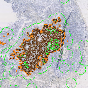
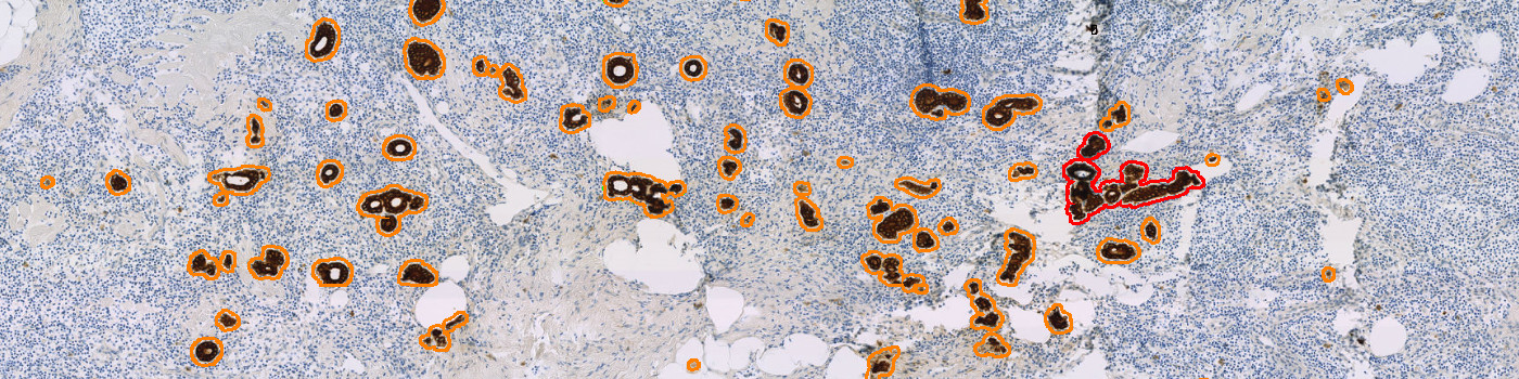
PCK positive tumor cells are identified in more detail (shown in orange).
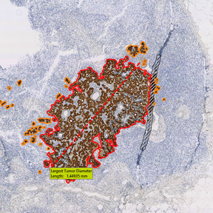
Largest tumor is identified, and the diameter is measured (shown in red).
Quantitative Output variables
The output variables obtained from this protocol are:
Workflow
The APP contains five protocols:
Step 1: Detect Brown: Roughly detects brown stained regions on a macroscopic level.
Step 2: Eliminate folds: Discards folds and similar artefacts from further analysis.
Step 3: Detect Tumor: Identifies PCK positive cells on a microscopic level.
Step 4: Calculate Largest: Finds the largest tumor and performs calculations.
Step 5: Detailed Outline: Simple post-processing to improve outline detail.
The protocols can be run separately or as one using the APP sequence functionality in VIS.
Methods
PCK positive cells are identified based on their strong red/blue contrast and color intensity. Very dark pixels are labeled PCK positive based on the surrounding brown hue and black artefacts such as carbon are eliminated. In case of positive spindle shaped cells (as seen with reticular and dendritic cells when using CAM5.2), the APP identifies those cells based on their low solidity and looks for similar cells dispersed in the neighborhood. If groups of low-solidity cells that are not densely grouped are found, they are labeled as non-tumor and excluded.
Staining Protocol
There is no staining protocol available.
Keywords
Lymph node, screening, metastasis, PCK, cytokeratin, immunohistochemistry, image analysis, breast cancer.
References
There are currently no references.