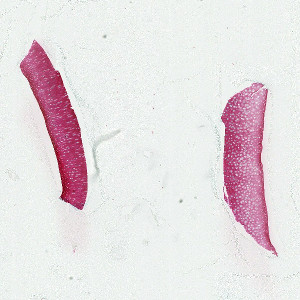
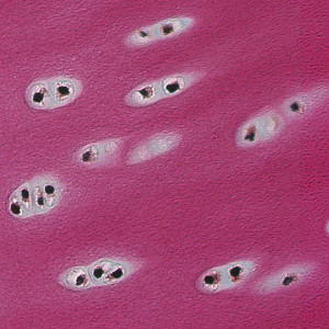
Two pieces of cartilage stained with Erlich Triacid.
#10054
In order to test the efficacy of different substances on the development of Osteoarthritis (OA) it is necessary to have a physiological relevant test system which includes not only chondrocytes, but cartilage matrix as well. This can be achieved using a cell culture like pellet culture or 3d culture, but the most physiological relevant test systems are cartilage explants. The explants can e.g. be derived from bovine or pig femur, tibia or metacarpus joint. To mimic diseased cartilage the explants are stimulated with cytokines or treated with single impact loading.
The model is suitable for studying basic pathophysiological aspects of OA and for the pharmaceutical development of disease-modifying drugs.
The severity of the cartilage destruction can best be demonstrated by histological processing of the explants by staining with Safranin-O-Fast Green or Toluidin Blue. The most important histopathological hallmarks in explants are decrease in chondrocyte number and loss of proteoglycan staining.
Traditionally, the evaluation of the severity of cartilage damage is done by subjective semi-quantitative grading of the different histopathological features using defined scoring systems. However, even with ‘blinding’ of the observers and sample randomization, this methodology is prone to bias, suffers from low sensitivity to change and requires considerable experience in histopathology. On the other hand, quantitative digital histomorphometry of cartilage destruction can offer an objective, less time consuming and more sensitive assessment of OA histopathology.
This APP can be used for quantifying profile density of chondrocytes in cartilage explants from bovine joints and is of relevance for different OA models, concerning pro anabolic or anti-catabolic factors. To answer this specific question a Ehrlich Triacid staining was choosen, because it allows the best differentiation between cartilage, bone and nuclei. The APP has been verified in treatment studies.
Two pieces of cartilage stained with Erlich Triacid.
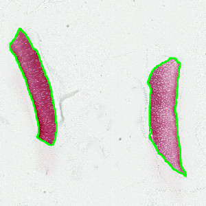
The cartilage identified as ROIs for subsequent analysis.
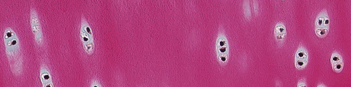
Closeup of the cartilage showing multiple chondrocytes.
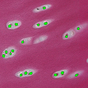
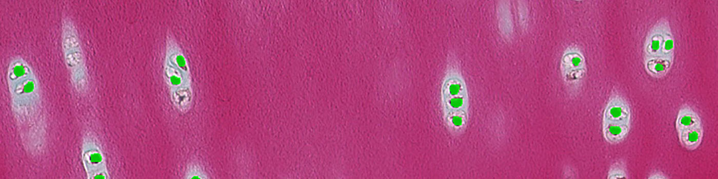
Segmentation of the image, separating the chondrocytes from the cartilage.
Quantitative Output variables
The output variables obtained from this APP include:
Methods
This APP can be used on virtual slides with several sections on each slide. The first step is the tissue detection on the slides. Followed by marking the tissue pieces and allocate them to ROI 1 to 4.
In a second step the cartilage area and the number of nuclei profiles can be obtained for calculation of the profile density.
Additional information
This APP was developed for, and validated by, Dr. Sven Lindemann, Simone Hartl, Merck KGaA, Osteoarthritis, General Research and Early Development.
Keywords
Osteoarthritis, Explant, Bovine, knee, cartilage, Ehrlich Triacid staining, quantitative, digital pathology, image analysis, bone density, chondrocyte.