Resources / Comparison of digital and computational algorithms for quantifying human epidermal growth factor receptor 2 (HER2) protein expression in metastatic breast cancer (mBC) from clinical samples
Insight
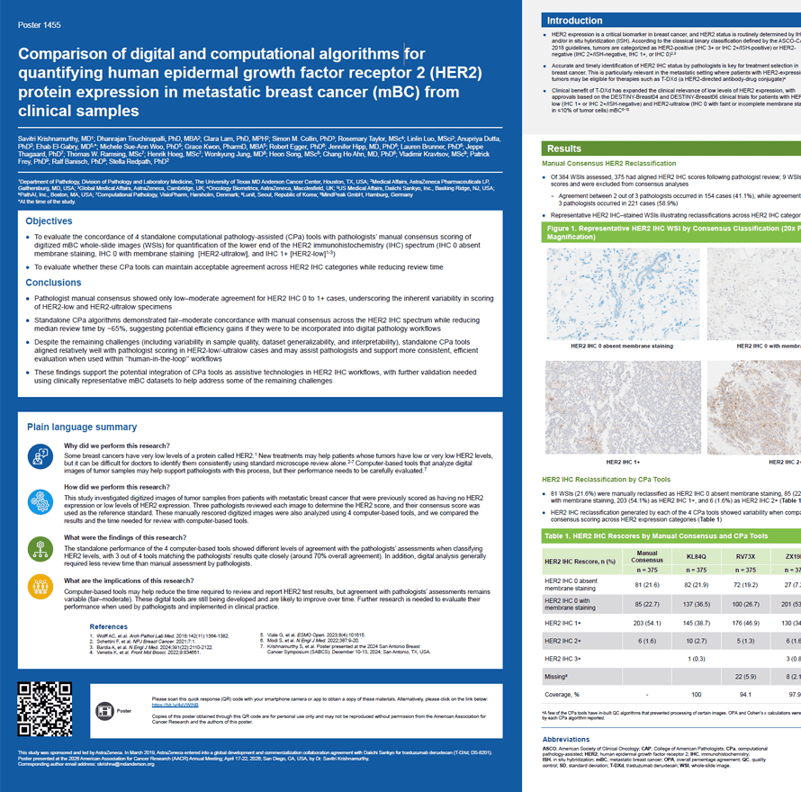
Comparison of digital and computational algorithms for quantifying human epidermal growth factor receptor 2 (HER2) protein expression in metastatic breast cancer (mBC) from clinical samples
Presented at AACR 2026
Description
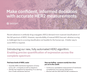
- HER2 expression is a critical biomarker in breast cancer, and HER2 status is routinely determined by IHC and/or in situ hybridization (ISH). According to the classical binary classification defined by the ASCO-CAP 2018 guidelines, tumors are categorized as HER2-positive (IHC 3+ or IHC 2+/ISH-positive) or HER2-negative (IHC 2+/ISH-negative, IHC 1+, or IHC 0)
- Accurate and timely identification of HER2 IHC status by pathologists is key for treatment selection in breast cancer. This is particularly relevant in the metastatic setting where patients with HER2-expressing tumors may be eligible for therapies such as T-DXd (a HER2-directed antibody-drug conjugate)
- Clinical benefit of T-DXd has expanded the clinical relevance of low levels of HER2 expression, with approvals based on the DESTINY-Breast04 and DESTINY-Breast06 clinical trials for patients with HER2-low (IHC 1+ or IHC 2+/ISH-negative) and HER2-ultralow (IHC 0 with faint or incomplete membrane staining in ≤10% of tumor cells) mBC
- Analyses of mBC samples originally scored as HER2 IHC 0 and IHC 1+ (HER2-negative) have showed that a substantial proportion of tumors may be reclassified as HER2-ultralow or HER2-low, and that inter-and intra-observer concordance among pathologists is variable, particularly at the lower levels of the HER2 IHC expression spectrum
- As the clinical relevance of HER2-low and HER2-ultralow has increased, accurate identification of very low HER2 expression has become increasingly important. Supporting pathologists’ confidence in interpreting these cases highlights the value for standardized protocols, additional training, and digital tools to support HER2 IHC scoring decisions
- Advances in the analysis of WSIs have transformed breast pathology by enabling workflows that support computational algorithms; CPa tools can help enhance diagnostic precision, reproducibility, and clinical decision-making
- This study builds on prior real-world findings11 of manual scoring by 3 pathologists and compares 4 standalone CPa tools versus pathologist-derived consensus scoring
Authors and institutions
Savitri Krishnamurthy, MD1, Dhanrajan Tiruchinapalli, PhD, MBA2, Clara Lam, PhD, MPH2, Simon M. Collin, PhD3, Rosemary Taylor, MSc4, Linlin Luo, MSci2, Anupriya Dutta, PhD2, Ehab El-Gabry, MD5*, Michele Sue-Ann Woo, PhD5, Grace Kwon, PharmD, MBA5, Robert Egger, PhD6, Jennifer Hipp, MD, PhD6, Lauren Brunner, Ph6, Jeppe Thagaard, PhD7, Thomas W. Ramsing, MSc7, Henrik Hoeg, MSc7, Wonkyung Jung, MD8, Heon Song, MSc8, Chang Ho Ahn, MD, PhD8, Vladimir Kravtsov, MSc9, Patrick Frey, PhD9, Ralf Banisch, PhD9, Stella Redpath, PhD2
- Department of Pathology, Division of Pathology and Laboratory Medicine, The University of Texas MD Anderson Cancer Center, Houston, TX, USA
- Medical Affairs, AstraZeneca Pharmaceuticals LP, Gaithersburg, MD, USA
- Global Medical Affairs, AstraZeneca, Cambridge, UK
- Oncology Biometrics, AstraZeneca, Macclesfield, UK
- US Medical Affairs, Daiichi Sankyo, Inc., Basking Ridge, NJ, USA
- PathAI, Inc., Boston, MA, USA
- Computational Pathology, Visiopharm, Hørsholm, Denmark
- Lunit, Seoul, Republic of Korea
- MindPeak GmbH, Hamburg, Germany
*At the time of the study.