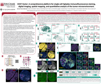
David Mason from Visiopharm spoke at Lunaphore’s Spatial Biology Week on an automated image analysis pipeline for high plex sequential immunofluorescent images. The challenges with this type of data include multiple platforms, unsatisfactory cell detection using traditional intensity-based approaches, and lack of confidence in results. David will use Lunaphore’s data sets, including a 22 Plex plus DPI tissue microarray, to demonstrate how Visiopharm can address these challenges in analyzing complex data sets.
-
- A guided bi-directional workflow designed specifically for setting phenotypes with continuous quality control and result review.
-
- Powerful pre-trained applications for detecting nuclei in multiplex immunofluorescence and imaging mass cytometry.
-
- User-friendly channel management tools for quality control of images and review of biomarker localization. Group your channels of interest into meaningful 7-color groups for easy switching between panels.
-
- An advanced interactive toolbox for data exploration and quality control, including t-SNE, scatter plots, and box plots.
David Mason, Senior Technical Specialist, Visiopharm
Dave Mason is a senior technical specialist in image analysis, supporting Visiopharm’s UK and European sales team. He has a background in cell biology and microbiology and has spent over a decade in academia, specializing in light microscopy and digital image analysis.